


Home Sample Collection

Highly Trained Staff & Doctors

Same Day Reports Over Email

Zero Contamination QualiCare Kit


Ultrasound, often referred to as a sonogram, is a procedure in which high-frequency sound waves are used to capture images of internal organs, tissues, and other structures in the human body. This is a non-invasive method that helps collect certain information about an individual’s body. For this imaging test, a thin layer of gel is applied, and a small device called a transducer is moved over the area that needs to be examined. High-frequency sound waves that are sent into the body’s tissue send electrical signals back so that real-time images or videos can be captured. Apart from being used to diagnose several health conditions, ultrasound scans are suggested during pregnancy as well. These scans help monitor foetal growth and detect any kind of complications.
Why are Ultrasounds Used for Prenatal Screening?
Being an important part of prenatal screening, ultrasound scans are primarily performed to keep track of the health and growth of the foetus. From determining the viability of a pregnancy to predicting the due date, from measuring the levels of amniotic fluid to ruling out any suspected complications, there are several ways in which an ultrasound scan can help doctors get an overview of a pregnancy. There are different types of ultrasounds performed in pregnancy. For example, an NT scan in pregnancy helps estimate the risk of any chromosomal abnormality in the foetus. Similarly, a Doppler scan in pregnancy is performed to check the blood circulation in the baby, uterus, and placenta.
Although each scan has its own significance, the number of pregnancy ultrasounds one has to undergo can vary from one pregnant female to another, depending on factors like age, medical history, etc. This is the reason why pregnant women without any risks of complications are suggested fewer ultrasounds as compared to those with a high-risk pregnancy.
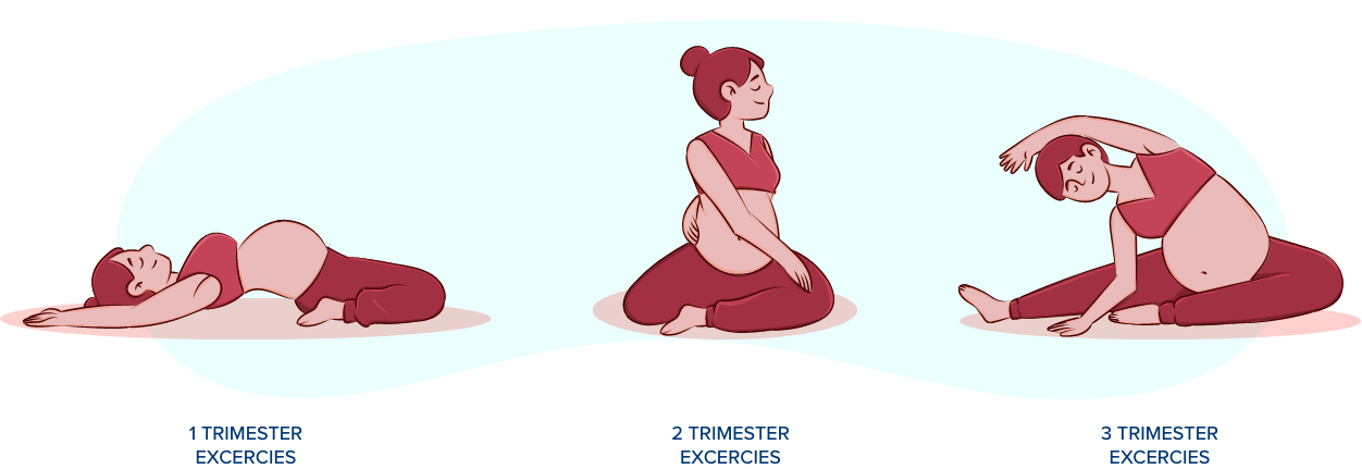
First Trimester Ultrasounds
After a positive pregnancy test, the next thing a doctor suggests is an ultrasound scan. Usually performed in the 6th week of pregnancy, this ultrasound helps not only confirm the pregnancy but also check the number of embryos and their positioning. Other than this, an early pregnancy scan also helps to calculate the gestational age and predict the due date for delivery.
Generally, two scans are performed in the first trimester, which are:
Viability Scan (6-9 weeks)
Early Morphology or Nuchal Translucency Scan (11-13 weeks)
Second Trimester Ultrasounds
The second trimester is a crucial period in terms of foetal growth, as this is when structures such as the spine, limbs, brain, and internal organs of the foetus start developing at a rapid rate. So, the scans suggested in this duration are focused on monitoring the health and growth of the baby. These ultrasound scans help the doctor determine if all the organs are developing the way they should and rule out any anomalies.
Here is a list of scans performed during the second trimester of pregnancy:
Anomaly Scan (18-21 weeks)
Growth Scan (26-18 weeks)
Third Trimester Ultrasounds
In the third trimester, ultrasound scans are performed to check things like movement of the limbs, stretching and flexing, breathing, opening and closing of hands, etc. All these things help ensure the baby is growing at a normal rate. As the pregnancy approaches its end, the doctor suggests checking the exact position of the baby and placenta, the amount of amniotic fluid, etc.
Mainly, two scans are performed during this trimester, which are:
Interval Growth Scan (28-32 weeks)
Term Scan (after 36th week)
However, in the case of a high-risk pregnancy, additional scans may be suggested to determine foetal well-being.
Get ready for your baby's arrival
.png)


 To reach our help desk call 7982100200